This is posts page. All of your news or blog posts should be listed on this very page, regardless the category they belong to. Posts page can have its own header image, owh crap, whatta nice, handy feature. WordPress is awesome.
In order to make Posts page “active”, select Settings > Reading from admin dashboard then be sure to tick select A static page radio button option. Use drop-down to navigate to this page.
We are SuKoKa members now
SuKoKa, Suomen Koirankasvattajat ry is an association of Finnish dog breeders founded in 1993
SuKoKa, Suomen Koirankasvattajat ry is an association of Finnish dog breeders founded in 1993, whose primary task is to promote responsible breeding work of purebred dogs and to act as a liaison and guardian of conscientious dog breeders and to contribute to the promotion of healthy dog breeding work and breeding.
SuKoKa, Suomen Koirankasvattajat ry is an association of Finnish dog breeders founded in 1993, whose primary... Continue
These is very important page. Because mom’s health = puppies health. So right feeding mom in pregnancy time and after birth mean a lot in my kennel.
Normally i feed all my dogs Royal Canin food. And i continue use these brend for pregnant females, mom’s and my puppies.
Just after mating i start give to future mom folic acid.
High maternal folic acid intake in the preconceptual period and early pregnancy greatly reduces the incidence of dermoid sinus in Rhodesian Ridgebacks just as it reduces the risk of related conditions in man.
In first week after mating i start give Raspberry Leaf. I find brend Animal Health from England and i order it from their main office.
Raspberry leaf provide symptomatic relief of problems associated with whelping and hey help in the prevention of pseudo-pregnancies in dogs and cats. The raspberry leaft tones the smooth muscle such as the uterus and helps to ensure a straightforward delivery
When future mom’s go in second part of pregnancy (after 30 days) i start give special food for pregnant females. Royal Canin HT42D Large Dog
Is your dog pregnant or in heat? Then it is especially important to support her with a good diet. Royal Canin HT42D Large Dog – Dog Food is specially formulated for pregnant and lactating bitches with an adult body weight starting at 10 kg. This dog food gives your dog just a little extra support during the first stage of the reproductive cycle. This phase consists of the 1st day of heat up to, and including, the 42nd day of gestation. This dog food ensures that your dog gets the right amount of energy and that the digestion is supported. Furthermore, these chunks contain delicious aromas, which will certainly make your dog love it!
In 10 days before birth and 10 days after birth i give special vitamins from Animal Health Easy Whelp.
Many problems can be encountered both at and after whelping including: Eclampsia, Mastitis, Metritis, Puppy mortality (fading puppy syndrome), Prolonged whelping, Bacterial diseases and many more
If whelping is slowed down, infection of the uterus is more likely to occur and increases the risk of still-births and weak puppies.
If we were able to encourage the ideal whelping, the following would be of high priority:
1) More rapid whelping. 2) Less disease. 3) Higher percentage of strong, viable puppies. 4) A less excitable/restless bitch-more settles and keen to feed and accept her puppies. 5) Rapid uterine recovery after whelping-less metritis, less discharge and more time for the uterus to ‘clean up’ Easy-Whelp is a feed supplement to aid normal whelping and lactation in bitches and to increase puppy viability.
It is a complete 20 day course which is added to the bitches’ feed 10 days before expected whelping and continued for 10 days after whelping. Easy-Whelp includes a very specific form of calcium which is the most readily available source of calcium by mouth.
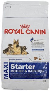
After female birth puppies her food changed on Royal Canin Maxi Starter Mother and Babydog
Complete feed for dogs – For the large breed bitch (from 26 to 44 kg) and her puppies: bitch at the end of gestation and during lactation – Weaning puppies up to 2 months old.
These is main food what i normally give to my females after mating and during all pregnancy. But it is no all what get my females. I give meat, milk products (Kefir, Quark, Yogurt…), Quail eggs, vegetables, fruits, berries… Some females has strong toxicosis during pregnancy and they want eat something special and i always try find what exactly female want eat.
These is very important page. Because mom’s health = puppies health. So right feeding mom in... Continue
A Role for Folic Acid in the Prevention of Dermoid Sinus in the Rhodesian Ridgeback
Dr John G. Roberts & Felicity A. Nicholls-Grzemski
Flinders Medical Centre & University of Adelaide South Australia
Dermoid Sinus (“sinus”), which can be single or multiple, was first reported in a Rhodesian Ridgeback (RR) in 1932. It has been the bane of RR breeders since that time and is widely believed to occur in approximately 10% of puppies. The true evidence has been obscured by the lack of a confidential reporting system and the reticence of many breeders to acknowledge its occurrence in their stock. Reputable Breeders seek early diagnosis of puppies in the first weeks of life and euthanase, or, on rare occasions, have the sinus surgically removed and the dog sterilised. Failure to remove a sinus leads later to cyst formation and potentially fatal infection.
Dermoid sinus is categorised as a “neural tube fusion defect (NTD)” by embryologists. When the neural groove, which runs longitudinally along what will be the back of the developing embryo, deepens and joins to form the new spinal cord and spinal column, the skin of the back also fuses in the midline and the spinal column and skin become completely separated. Partial failure of this process leads to dermoid sinus in the RR and human defects ranging from “dermal sinus” (an identical condition) to spina bifida occulta (hidden) to spina bifida cystica with serious paralysis. RRs do not exhibit this extreme form of NTD.
The neural tube fusion and separation process is complete in humans by day 35 post-conception, i.e. very early in the normal 280 day gestation period. As dogs have a 63 day gestation, this fusion (or failure of fusion) occurs within the first 2 or 3 weeks after conception. This very early establishment of any fusion defect is critical when considering possible preventative strategies. It is over and done with before pregnancy can be diagnosed reliably, certainly in the dog.
In July 1991 an immensely important research paper was published in The Lancet (UK). This was “Prevention of Neural Tube Defects: Results of the Medical / Research Council Vitamin Study”. This paper reported results which have changed world thinking on dietary folic acid (folate) – a non-toxic water soluble vitamin -supplementation before and during pregnancy. The study was based on earlier observations that lack of a nutritious balanced diet in the mother appeared to predispose babies to NTDs.
A large multi-country prospective trial was established. Women who had previously had a child with an NTD were recruited under strictly controlled conditions. These women were given one of four treatments commenced before conception. These were Vitamins A, D, B1 – B6, C, nicotinamide or Vitamins as above + folic acid 4 mg or Folic acid 4 mg or Iron/Calcium each taken daily until at least three months of pregnancy. Maternal diet was not controlled. A definite result, i.e. whether the foetus / baby was definitely affected or not affected with an NTD, was obtained in 1195 pregnancies. The study was then stopped because the results were so totally clear that the study could not ethically continue.
The incidence of neural tube defects in pregnancies of mothers in either of the two folic acid groups was reduced by 72% compared to the incidence in the non-folic acid groups. This information has revolutionised perinatal nutrition guidelines. The USA has recently mandated folic acid enrichment of basic grain-based foodstuffs.
This led one of us (JR) to wonder why this unique advance in human medicine might not be applied to a similar condition (sinus) in the RR.
The collaboration of a research pharmacologist / toxicologist (F.N-G) was arranged. After approval by the RRCSA a prospective open study was promoted personally, in lectures and in dog club journals. A detailed questionnaire was forwarded to interested parties. It was left to respondents whether or not they supplemented their bitches’ diets with folic acid 2.5 to 5 mg / day from the time of mating if not earlier. Details of average diet and history of sinus in the breeding pairs’ birth litters were inquired of in detail. Respondents reported, inter alia, litter size, sex of puppies, occurrence of dermoid sinus and any other congenital abnormalities.
RESULTS To February 1996, reports of 51 litters comprising 429 puppies had been received. Subsequent reports will be included in our definitive paper for publication.
Bitches on a “normal” diet (meat, bones, scraps, commercial dog food) produced 25 litters (213 puppies), bitches receiving a high folate (high vegetable) diet calculated to yield at least 200 mcg folate / day had 11 litters (93 puppies) while folic acid tablet supplemented bitches (15) had 123 puppies.
“Risk” of sinus, based on sire and dam’s birth litter sinus incidence, was rated as “high” (sinus in the birth litters of both), “medium” if one parents birth litter was affected, “low” if neither’s birth litter had sinus and “unknown”. There was no correlation between perceived risk and the occurrence of sinus in the puppies reported in this study.
The incidence of sinus in the “normal (low folate) diet group was 16.0% The two high folate groups (diet or tablets) combined showed an incidence of sinus of 4.2%. In the high folate diet group zero incidence of sinus was reported. The folic acid supplementation by tablet group had a sinus incidence of 7.3% (Table 1).
All the differences between the incidence of sinus in the low folate control group and that in the high folate groups are statistically significant (Wilcoxon rank-sum test, Mann Whitney U-test).
DISCUSSION The results clearly indicate that high folate levels in the diet of the breeding Rhodesian Ridgeback bitch lead to a significant and important reduction in the incidence of the NTD, dermoid sinus, in their progeny. This is analogous to the proven situation in man. A reduction of around 70% can be reasonably expected.
The small sample of high vegetable folate litters reported a zero incidence of sinus while litters receiving folic acid tablets showed a 55% reduction. Why might this difference exist? The answer is unlikely to relate to other vitamins in the vegetable diet as this was not the human experience. It is most likely due to the fact that high folate diet bitches received a continuous and automatic supply of folate during their adolescence, sexual maturity, mating and pregnancy. No one had to remember to start tablet administration or to continue it on a daily basis – all the very limited body stores of folate would be continually full as would the developing ova. The converse applied to the folic acid tablet supplementation group where the time of starting treatment in relation to the bitches cycle was sometimes not as recommended. Two bitches included in the folic acid tablet group were started on tablets at one week and ten days post mating (too late), while another bitch included in that group was commenced “on confirmation of pregnancy” (far too late). Such inclusions in the folic acid treatment group can only have reduced the observed 55% protective effect which would be expected to be higher in the face of timely or continuous supplementation.
CONCLUSION High maternal folic acid intake in the preconceptual period and early pregnancy greatly reduces the incidence of dermoid sinus in Rhodesian Ridgebacks just as it reduces the risk of related conditions in man. A strong case can be made for either a high folate diet or continuous folic acid supplementation of “normal” diets throughout the reproductive life of RR bitches.
A Role for Folic Acid in the Prevention of Dermoid Sinus in the Rhodesian Ridgeback Dr... Continue
On the 8th of july 2001, my third litter was born, out of Kwetu’s Springtime in Holland (Kana, Imp.USA) and my own Holmland Owl’s Rafiki Cali.
Cali is from a DS-free litter, in Kana’s litter there was 1 DS. Kana herself has been on a high folate diet throughout her entire life.
At first, the litter looked very promising, 6 ridged pups and only 1 faulty ridge (single crown). Unfortunatly, after a couple of days we discovered that 4 pups had a Dermoid Sinus, all in the neck area.
I was faced with a very difficult choice, whether to cull the pups or have them operated on.
I found there was very little information on DS surgery available and the few articles on the Internet were all quite old and very negative about the surgery.
After speaking with several breeders and owners of dogs that had a DS removed and a long talk with a very experienced surgeon, I learned that the surgery wasn’t in fact that traumatic and in 90 % of the cases a total succes! So I decided to give my pups a fair chance.
Since I was fortunate enough to have a long waiting list with potential puppy-buyers, I had no trouble finding the puppies a good home. I also kept one puppy myself.
The new owners were of course well informed about the DS, the surgery and the consequences of this genetic defect. They knew in advance that their puppy was not show or breed material, and had to be spayed/ neutered. All the pups were placed in wonderful homes, at the age of 8 weeks.
At the age of 12 weeks (the surgeon doesn’t like to put younger pups under aneasthesia) we had all 4 pups operated, in two days time. I was present at all procedures and able to see for myself all the DS’s being removed. Two of the pups (including my own) had a quite thick, long DS, all the way to the vertebra.
Fortunatly, they did not go into the spine. The other two were more superficial and easy to remove. One pup had two smaller DS’s, both in her neck. They were both easily removed. The pups came home with us a couple of hours after the surgery. Their recovery was amazing, that same evening we already had trouble keeping them quiet! They were not in any pain and recovered quickly from the aneasthesia.
The pups had to wear a neck-bandage for two weeks, to prevent serum built-up and could not play with other dogs for that time. In stead we took them for trips into town and walks on leash, so they wouldn’t miss out on any socialisation. They were even able to go to puppy-class as usual.
My own puppy had some swelling under the wound, but after removing the fluid once, it didn’t come back. The other 3 puppies didn’t have any complications.
After a couple of weeks, it was all over and the scars weren’t even visible anymore. The pups are now 6 months and no one can tell the diffenence between the operated pups and their siblings. I am very glad with the results and have never regretted my decision to let these puppies live. Their owners are very responsible people, who will have the dogs spayed/ neutered when they are old enough (in Europe, we don’t do early spays). The 2 pups that are not afflicted will also be spayed, as is their mother. I feel very strongly that dogs that come from a high DS incidence litter should not be bred.
The cost of the surgery was about 1600 Euro, payed by me of course. But seeing those happy, healthy pups and their proud owners is worth every cent.
Kiku 7 weeks old. She had a large DS in the neck, which was not clearly visible at first sight.
Kiku in a different light, from a different angle, we now see a “suspicious” spot, where the hairs seem te have a slightly different colour and structure. When you feel the spot, there seems te be a small “lump” under the skin.
When the skin is lifted, the DS is clearly visible, as a “thread” which runs down from the skin to the spinal cord. Also note the “dip” in the skin.
when the area is shaved, the exit of the DS is clearly visible. This particulair one has two exits!
a different puppy, with a smaller DS. There was absolutely nothing to see from the outside. But when she was shaved there was no doubt, the exit is there.
Kiku, prepared for surgery.
The DS is the white “thread” on top of the surgeons finger.
The DS has been separated from the surrounding tissues.
The neck bandages didn’t really bother the pups…
Some serum built up, despite the bandage. It had to be removed only once though.
This is the puppy that had 2 DS’s. She healed without any complications.
Note, this article is in no way meant as an advertisement for DS surgery. The decision whether to cull or operate is up to each individual breeder. I do believe however, that the decision should be made based on facts and information and not on the slightly one-sided information that has been available up to now.
Also, I cannot stress enough that ALL pups should be placed with responsible owners, that do not breed dogs that have any kind of genetic problem.
Anke Terbruggen – The Netherlands 20-01-2002
PS – pictures and text can be copied freely and without permission from the author.
Kiku 6 months old. There isn’t even a scar anymore.
On the 8th of july 2001, my third litter was born, out of Kwetu’s Springtime in... Continue
The Rhodesian Ridgeback is a modern breed of dog that originated in the late nineteenth century, by the crossing of indigenous Hottentot dogs with various European breeds introduced into the Cape by the early settlers.
The breed standard was established with the formation of the Rhodesian Ridgeback Club of Bulawayo in 1922. The main characteristic of the breed is, as its name implies – a ridged back, which is formed in the hair/coat along the top midline of the dog’s back. The ridge is formed by hair, which grows in the opposite direction to the hair of the surrounding coat.
Breeders of Ridgebacks are aware of a well-known defect which occurs in the breed, the Ridgeback “Cyst” or as it is more correctly named in the scientific terminology, the Dermoid Sinus. (Dermoid – arising from the skin, Sinus – a cavity or channel).
Dermoid Sinuses are narrow tube-like structures, which are derived from a skin defect. They penetrate from the skin surface to varying depths downward into the muscles and towards the spinal cord. They are situated in the midline of the neck and croup, which is in front and behind the area occupied by the ridge (Fig 1).
Fig 1: Areas marked X indicate the sites at which dermoid sinuses may develop.
This is the only known congenital defect that occurs regularly in the breed. (Congenital means that the defect is formed before birth). When considered as a defect in the dog family as a whole, Dermoid Sinuses occur only very rarely in dogs, other than Ridgebacks or Crossbred Ridgebacks. It must therefore be obvious that it is an inherited defect which has become widespread in the “blood lines” of the breed as a result of the early selective breeding of the original stock from which the Ridgebacks of today have been produced.
The incidence of the defect throughout the breed is not known, as the recording of the numbers of Dermoid Sinus affected pups in litters has not been done on a scale large enough to enable a statistical analysis to be carried out. In fact, the occurrence of Dermoid Sinus affected pups in the litters of breeders has been kept confidential, as most breeders feel that there is considerable stigma attached to dogs and bitches amongst whose offspring Dermoid Sinus affected puppies occur.
At this point I would like to state that with the present situation of breeding with selected outstanding dogs and bitches, no breeder without a program of progeny testing can be sure that his “blood line” is free from the hereditary Dermoid Sinus. (The hereditary aspects of the condition will be dealt with in part two of this article). Thus, every purchased Ridgeback may be considered a potential carrier of the condition.
The formation of Dermoid Sinus
To understand the way, in which a Dermoid Sinus is formed, it is necessary to have some idea of how the embryo develops from a single fertilized egg cell in the womb of the bitch. Dermoid Sinus is a congenital defect that arises from a defect in the development of the embryo of a puppy.
A fertilized egg resulting from a successful mating is a single simple cell. From this cell a puppy consisting of millions of specialized cells, which constitute the tissues and organs, must be formed in 63 days.
Fig 2: Early stages of cell division.
A: Single cell of fertilized egg.
B: 2 cell stage.
C: 4 cell stage.
D: 8 cell stage.
E: Multi-celled Spherical Mass, many cell divisions later.
This process in accomplished by a rapid increase in the number of cells by cell division. The fertilized egg (a single cell) divides into two cells and subsequent divisions each double the previous number of cells, so that the numbers very rapidly increase. In the ten successive divisions, 1042 cells are produced, and it can be seen that, by this means, the total number of cells is soon very large.
The next stage is the organization of the mass of cells produced to form a puppy. The organization process, which takes place for about the first three weeks of pregnancy, is called the embryonic development. When the embryo is fully developed, a complete miniature puppy is formed which now becomes known as a foetus. The next six weeks of pregnancy only results in the increase in size of the foetus to its normal birth size.
Dermoid Sinuses arise from a defect in the development of the embryo. Cell division gives rise to a spherical mass of cells. The outer layer of these cells will eventually become the skin of the puppy. Another part of the body also develops from this outer layer of cells. This is the brain and the spinal cord, which runs from the head to the base of the tail. The problem now is – how does the outer layer of cells give rise to the brain and the spinal cord?
This is accomplished by the formation of a long groove over half the surface of the spherical mass of cells. The groove deepens and then its outer edges close together giving rise to a tube-like structure. This tube-like structure which is later to become the brain and spinal cord, sinks deeper below the surface layer and becomes detached from it. This process is shown in Figure 3.
Fig 3: Schematic Formation of Brain and Spinal Cord
A: Formation of groove on surface of sphere of cells as seen from above groove.
B: Section through X-Y to show outer layer of cells folding inwards.
C: Deepening of the fold
A: Closing over of edges of the groove.
B: Closed.
C: Separation of outer layer (skin) from tubular structure (spinal cord).
Dermoid Sinuses occur when small areas of attachment between the outer layer of cells (the skin) and the tubular structure (later to become the brain and the spinal cord) remain.
In the puppy this defective separation of the embryological tissues is present as a thin tubular attachment extending from the skin of the midline of the top surface of the dog to the deeper tissues below, and as deep as the spinal cord in some cases.
The depth to which this tubular skin defect penetrates is the criterion used for the classification of four types of Dermoid Sinuses, shown in Figure 4 below.
Cross section through a dog’s neck Types of Dermoid Sinuses
TYPE I Penetrates below skin surface, its fatty tissue overlying the neck muscles.
TYPE II Penetrates into the muscles of the neck.
TYPE III Penetrates to supraspinous ligament, running over the top of the vertebrae.
TYPE IV Penetrates to the spinal cord between the vertebrae.
The significance of Dermoid Sinud
The detrimental effects of Dermoid Sinus are not just concerned with the fact that a visible anatomical defect is apparent in affected animals, but rather the complications which can arise as a result of a Dermoid Sinus becoming infected with bacteria.
The narrow tube of skin which descends below the skin surface is lined with all the normal skin structures and of special significance are : hair; sweat; and oil glands.
The thin central cavity, which runs down the Dermoid Sinus, becomes filled in time with hair, skin oil and skin scales. The contents usually become an ideal medium where bacteria, which are normally present on the skin, may grow. They gain access to the material through the small pore-like opening at the point of attachment of the Dermoid Sinus on the skin surface.
The accumulated skin secretion undergoes a process of putrefaction and the skin barrier of the Sinus walls breaks down and bacteria invade the tissues deep below the skin surface. This usually results in the formation of an abscess, which eventually ruptures to the outside and drains as a chronically discharging purulent wound.
Extensive surgical and medical treatment may be necessary to clear up such a complication and in some cases septic dermoid sinus may be unresponsive to treatment.
If a Dermoid Sinus is recognized in a dog before it becomes septic, it can be removed surgically, with a good chance that no further complications will occur. In most cases, however, owners of animals are not aware of the presence of a Dermoid Sinus and shortly thereafter sepsis almost always sets in. Subsequently, the owners are obliged to obtain veterinary treatment to resolve the distressing complications. This may be costly to the dog owner and embarrassing to the breeder when it is pointed out that he has sold a dog with a latent defect.
Diagnosis of Dermoid Sinus
In the puppy, Dermoid Sinus can be detected by raising the skin in a longitudinal fold along the top midline in the area in which Dermoid Sinuses are known to occur (i.e. in front of and behind the ridge). If the skin fold is raised with one hand and the skin allowed to slip back and forward between the thumb and forefinger of the other hand the presence of the Sinus can be felt as a thin cord-like structure between the two layers of skin (Fig 5)
Figure 5: Diagnosis of Dermoid Sinus
Feeling for presence of Dermoid Sinus by sliding longitudinal fold of skin between index finger and thumb. Raising the skin fold in this way tenses the tissues and a Dermoid Sinus will be pulled tautly between its skin attachment in the top midline and its attachment in the muscles below.
The diagnosis can be confirmed by shaving the hair from the skin over the point at which the Dermoid Sinus is attached. A small pore like opening in the skin from which a small tuft of hair protrudes is usually seen. This is the opening of the Dermoid Sinus on the skin surface. The older the puppy, the thicker the Sinus will be and the more easily it may be recognized.
It must be realized, however, that the recognition of a Dermoid Sinus in puppies may not always be as easy a procedure as the above description may suggest. If it is missed, a Dermoid Sinus may lie dormant for years before it comes to the notice of an owner by becoming septic. If in doubt the professional assistance of a Veterinarian should be obtained.
An excellent site showing the dermoid sinus in pups and the operation undertaken to remove those sinuses can be found at